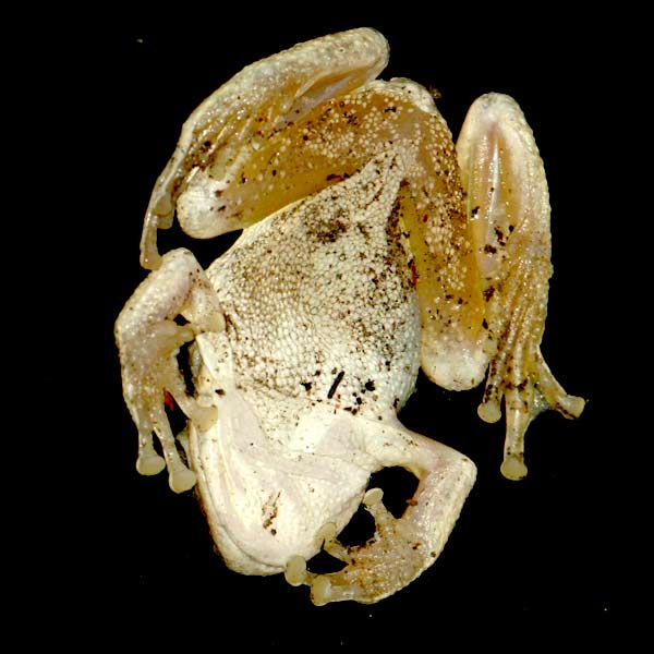
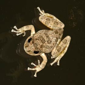
Tree frogs in trees are just fine, but tree frogs on plate-glass windows are better – because then you get to see their slightly icky fascinating undersides.
I must admit, what struck me most when this guy landed – ‘thunk’ – out of a fig tree onto our window in Los Angeles, was how much his (her?) legs looked like raw chicken. I’ve always shied away from those cuisses de grenouille opportunities, but I know the meat is often compared to chicken. Why frogs and chickens developed that way is an interesting question, but not for today’s post.
Rather, now I’m back in the UK , where the frogs are less acrobatic, I’ve tried to figure out how our unexpected visitor managed to cling on.
Wet Adhesion
To understand that for the West Indian tree frog, Oseopilus septentrionalis, researchers Hanna and Barnes1 used active and anaesthetised frogs in experiments that measured the forces they apply walking up vertical surfaces, the angle at which they drop off a gradually inclined surface, and the shear force experienced by an individual toe when the surface it’s attached to is suddenly slid from under it.
The experiments involved placing frogs on a variety of strain-gauge instrumented platforms and surfaces, and making videos of frogs placed on runways and rotating discs of transparent perspex.
The researchers concluded that the primary mechanism tree frogs use to get a grip is exactly the same as that which keeps a sheet of wet paper stuck to the side of a glass: wet adhesion.
Wet adhesion combines a mix of viscous and surface tension forces, both of which require liquid and, in the case of surface tension, an air-liquid interface. In the tree frog, that liquid takes the form of mucous – wait for it – pumped out the ends of its toes.
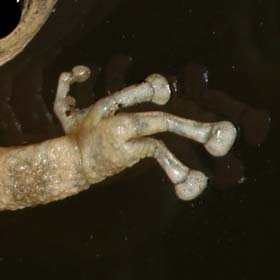
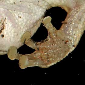
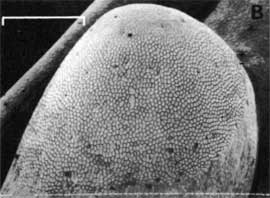
The mucous appears from perfectly smooth-looking toe pads that are actually covered in thousands of peg-like cells between which mucous flows from glands. I can see something going on in my own photographs, but the structure is clear in the SEM picture below.
Hanna and Barnes’s also looked at how tree frogs release themselves to move. The frogs peeled rather than pulled their feet off surfaces, the peeling force engaging automatically in forward movement, but not in reverse or when the belly skin of the frog made contact with the surface.
Frogs placed on a slowly rotating vertical disc reorientated themselves to avoid facing downwards – presumably because of the involuntary forward travel or detachment that would induce. At first sight then, my Pseudacris cadaverina appears to defy that rule – because he’s clearly inverted in one of the pictures; but that could be explained by the extra adhesion he’s getting from the inner thigh area – corresponding to the aforementioned belly skin. What’s more, the authors point out that toe pads have developed independently several times in tree frogs, so the observed peeling mechanism may be peculiar to Oseopilus septentrionalis.
I would have liked to spend more time with this little guy, but after about ten minutes I looked up and he’d gone – probably back into the fig tree. But for a while there he sure provided a level of interest, conversation, and intrigue way out of proportion to his size. Ribbit.
References
1. Adhesion and detachment of the toe pads of tree frogs. Gavin Hanna, W.John Barnes. Journal of Experimental Biology 155, 103-125 (1991)
